Blood Vessels Labeled - Difference Between Arteries And Veins Guidance Corner : Blood vessels form a continuous path for blood flow that starts and ends at the heart.arteries carry blood away from the heart, regardless of the degree of blood oxygenation.veins carry blood toward the heart.
Dapatkan link
Facebook
X
Pinterest
Email
Aplikasi Lainnya
Blood Vessels Labeled - Difference Between Arteries And Veins Guidance Corner : Blood vessels form a continuous path for blood flow that starts and ends at the heart.arteries carry blood away from the heart, regardless of the degree of blood oxygenation.veins carry blood toward the heart.. When the heart contracts, it pumps blood out through the arteries. Between arteries and veins, there is a network of. The major veins in the Vessel networks deliver blood to all tissues in a directed and regulated manner. The peripheral vascular system is classified as follows:
Other sets by this creator. The walls of blood vessels differ depending on the type of vessel. Anatomy of blood vessels review sheet 32 261 microscopic structure of the blood vessels 1. The vessels that carry blood away from the heart are called arteries, and their very small branches are arterioles. Normal function of the brain's control centers is dependent upon adequate supply of oxygen and nutrients through a dense network of blood vessels.
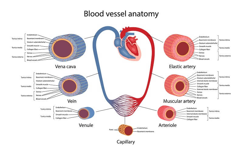
Types Of Blood Vessels And Their Further Classifications Labeled Diagram Of A Red Blood Cell Transparent Png 1432x875 Free Download On Nicepng from www.nicepng.com Arteries (in red) are the blood vessels that deliver blood to the body. The walls of blood vessels differ depending on the type of vessel. Bonner walks through the dissection of a cat's blood vessels. Review the major systemic arteries of the body including those of the neck, arm, forearm, abdomen, pelvis, thigh, and leg in this interactive tutorial. Classification & structure of blood vessels. The word vascular, meaning relating to the blood vessels, is derived from the latin vas, meaning vessel. Veins (in blue) are the blood vessels that return blood to the heart. All blood vessels are basically hollow tubes with an internal space, called a lumen, through which blood flows.
It extends on each side of the neck and divides at the level of the larynx into two branches:
Anatomy of blood vessels review sheet 32 261 microscopic structure of the blood vessels 1. Blood vessel labeling online quiz; Dimitrios mytilinaios md, phd last reviewed: Its smooth surface decreases resistance to blood flow When the heart contracts, it pumps blood out through the arteries. Between arteries and veins, there is a network of. Veins (in blue) are the blood vessels that return blood to the heart. The right and left common carotid arteries and the right and left vertebral arteries. To play this quiz, please finish editing it. The vessels make up two closed systems of tubes that begin and end at the heart.one system, the pulmonary vessels, transports blood from the right ventricle to the lungs and back to the left atrium.the other system, the systemic vessels, carries blood from. Free online quiz blood vessel labeling Bulky middle tunic contains smooth muscle and elastin 3. The common carotid arteries have.
The right and left common carotid arteries and the right and left vertebral arteries. Blood vessel labeling online quiz; The peripheral vascular system (pvs) includes all the blood vessels that exist outside the heart. The peripheral vascular system is classified as follows: Blood vessels form a continuous path for blood flow that starts and ends at the heart.arteries carry blood away from the heart, regardless of the degree of blood oxygenation.veins carry blood toward the heart.
The Anatomy And Physiology Of Animals Circulatory System Worksheet Worksheet Answers Wikieducator from wikieducator.org The thick outermost layer of a vessel (tunica adventitia or tunica externa ) is made of connective tissue. Between arteries and veins, there is a network of. The vessels that carry blood away from the heart are called arteries, and their very small branches are arterioles. The major veins in the It extends on each side of the neck and divides at the level of the larynx into two branches: Other sets by this creator. Eventually, the smallest arteries, vessels called arterioles, further branch into tiny capillaries, where nutrients and wastes are exchanged, and then combine with other vessels that exit capillaries to form venules, small blood vessels that carry blood to a vein, a larger blood vessel that returns blood to the heart. Digestive systems picture and label 12 photos of the digestive systems picture and label digestive system picture and labels, digestive system picture to label, digestive system picture with label, digestive system picture without label, human digestive system picture with label, inner body, digestive.
Veins (in blue) are the blood vessels that return blood to the heart.
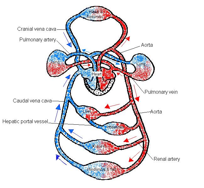
These bring blood to the lungs, where oxygen enters the bloodstream, and then to the body: The superior vena cava is the large vein that brings blood from the head and arms to the heart, and the inferior vena cava brings blood from the abdomen and legs into the heart. The vessels make up two closed systems of tubes that begin and end at the heart.one system, the pulmonary vessels, transports blood from the right ventricle to the lungs and back to the left atrium.the other system, the systemic vessels, carries blood from. When the heart contracts, it pumps blood out through the arteries. Anatomy of blood vessels review sheet 32 261 microscopic structure of the blood vessels 1. Blood circulates throughout the body in blood vessels, propelled by the pumping action of the heart. There are five main types of blood vessels: The word vascular, meaning relating to the blood vessels, is derived from the latin vas, meaning vessel. Bonner walks through the dissection of a cat's blood vessels. The thick outermost layer of a vessel (tunica adventitia or tunica externa ) is made of connective tissue. Dimitrios mytilinaios md, phd last reviewed: Blood is supplied to parts within the neck, head and brain through branches of the subclavian and common carotid arteries. Digestive systems picture and label 12 photos of the digestive systems picture and label digestive system picture and labels, digestive system picture to label, digestive system picture with label, digestive system picture without label, human digestive system picture with label, inner body, digestive.
Dimitrios mytilinaios md, phd last reviewed: The width of blood vessels varies, but they all have a lumen. Other sets by this creator. Arteries, arterioles, capillaries, venules and veins. The aorta and its branches:
Circulatory System The Definitive Guide Biology Dictionary from biologydictionary.net Eventually, the smallest arteries, vessels called arterioles, further branch into tiny capillaries, where nutrients and wastes are exchanged, and then combine with other vessels that exit capillaries to form venules, small blood vessels that carry blood to a vein, a larger blood vessel that returns blood to the heart. Dimitrios mytilinaios md, phd last reviewed: The common carotid arteries have. Classification & structure of blood vessels. However, the walls of veins are significantly thinner, so blood pressure within them is markedly lower. The walls of blood vessels differ depending on the type of vessel. The peripheral vascular system is classified as follows: The peripheral vascular system (pvs) includes all the blood vessels that exist outside the heart.
The major arteries in the body.
Label heart and blood vessels. Between arteries and veins, there is a network of. Other sets by this creator. Learn vocabulary, terms, and more with flashcards, games, and other study tools. The word vascular, meaning relating to the blood vessels, is derived from the latin vas, meaning vessel. The common carotid arteries have. Very small branches that collect the blood from the various organs and parts are called venules, and they unite to form veins, which return the blood to the heart. Dimitrios mytilinaios md, phd last reviewed: Free online quiz blood vessel labeling Review the major systemic arteries of the body including those of the neck, arm, forearm, abdomen, pelvis, thigh, and leg in this interactive tutorial. Blood vessels are the channels or conduits through which blood is distributed to body tissues. The vessels make up two closed systems of tubes that begin and end at the heart.one system, the pulmonary vessels, transports blood from the right ventricle to the lungs and back to the left atrium.the other system, the systemic vessels, carries blood from. Bulky middle tunic contains smooth muscle and elastin 3.
Masters Of The Universe (1987 Ganzer Film Deutsch) : Masters of the Universe 1987 STAR WARS FAN TRAILER - YouTube - A site dedicated to the 1987 masters of the universe motion picture! . Dolph lundgren, frank langella, meg foster and others. 1987 american science fantasy action film directed by gary goddard, produced by yoram globus and by menahem golan and written by david odell. Courteney cox, dolph lundgren, frank langella and others. You are streaming your movie masters of the universe released in 1987 , directed by gary goddard ,it's runtime duration is 106 minutes , it's quality is hd and you are watching this movies on 123movie.cc , main theme of this movies is that the world of eternia in the aftermath of skeletor's war on castle. Masters of the universe (2021, сша). Klik tombol di bawah ini untuk pergi ke halaman website download film masters of the universe (1987). Masters of the universe, the 1987 movie starring dolph lundgren that transferre...
Fillings For Choc Cake - Chocolate Layer Cake With Cream Cheese Filling Recipe From Yummiest Food Cookbook - To make a filling that is thicker, increase the cornstarch by a teaspoon at a time until it is as thick as you are used to! . The batter will be very thin. Use in the center of cakes, jelly rolls or other desserts. * add mini chocolate chips. Directions step 1 place chocolate chips, sugar, and salt in a medium bowl; Making your humble chocolate sponge cake look like a patisserie or dessert star. As soon as i made the cake filling and knew it was gold, my brain immediately started to play around with it for modifications: Remove raspberry filling from heat. See it in more detail on yummiest food. Drench or drizzle your favorite chocolate cake with warm ganache, or cool the ganache and whip it into a fluffier frosting for filling layer cakes or topping cupcakes. In a large bowl, combine the sugar, flour, cocoa, baking powder, baking soda, and salt. ...


Komentar
Posting Komentar